Anatomy Of Chest Area - Sternum - Wikipedia - Anatomy is to physiology as geography is to history:. Structures that pass through this area can be. This chapter is an abbreviated review of thoracic anatomy as seen on chest radiographs the retrocrural space (aortic hiatus) is the space bounded by the diaphragmatic crura and the spine. There the heart beats an average of 72 times a minute and circulates up to 2000 gallons of blood a day. The stomach is located inside the abdominal cavity in a small area called the bed of the stomach, onto which the stomach lies when the body is in a supine position, or. A good radiologist knows the anatomy, so don't skip this chapter!
Chest , chests , thorace , thoraces , thorax , thorax , chest region , chest , chest , chest region , area thoracic , chest and upper back , thoracic region , thoracic area , thoraces , regions thoracic , thoracics , thorax , thoracic , thoracic , thoracic structure , thoracic (qualifier value) , thoracic. Ct anatomy of the chest, axial reconstruction. Hilar lymph nodes are not visible unless abnormal. The stomach is located inside the abdominal cavity in a small area called the bed of the stomach, onto which the stomach lies when the body is in a supine position, or. The epidermis is the outermost layer that provides a protective, waterproof seal over the body.
A Quick Guide to Chest X-Rays - IVLine from 4.bp.blogspot.com There the heart beats an average of 72 times a minute and circulates up to 2000 gallons of blood a day. The chest exam is performed more frequently than any other exam in the imaging department. It consists of four parts, two curvatures and receives its blood supply mainly from the celiac trunk. The electrical impulse then travels to an area of cells at the bottom of the right atrium, between the atria and ventricles, called the atrioventricular, or av, node. We have other charts available that map these areas on hands and feet. 1, inferior lobe of right lung. Hilar lymph nodes are not visible unless abnormal. The area where the esophagus joins the stomach is called the gastroesophageal (ge) junction.
The interpretation of a chest film requires the understanding of basic principles.
This mri chest (thorax) axial cross sectional anatomy tool is absolutely free to use. It consists of four parts, two curvatures and receives its blood supply mainly from the celiac trunk. The function of the lungs is to oxygenate blood. A collection of anatomy notes covering the key anatomy concepts that medical students need to learn. Anatomy of the chest and the lungs: Learn about the anatomy and physiology of the stomach. Anatomy is to physiology as geography is to history: The chest anatomy includes the pectoralis major, pectoralis minor & serratus anterior. This chapter is an abbreviated review of thoracic anatomy as seen on chest radiographs the retrocrural space (aortic hiatus) is the space bounded by the diaphragmatic crura and the spine. Learn about each muscle, their locations & functional anatomy. Indications for mri •a chest mri provides detailed pictures of tissues within the chest area. It describes the theatre of events. There the heart beats an average of 72 times a minute and circulates up to 2000 gallons of blood a day.
In fact every radiologist and pulmonary physician should be an expert in chest film reading. Anatomy of the chest and the lungs: They are located in the chest, either side of the mediastinum. Indications for mri •a chest mri provides detailed pictures of tissues within the chest area. This chapter is an abbreviated review of thoracic anatomy as seen on chest radiographs the retrocrural space (aortic hiatus) is the space bounded by the diaphragmatic crura and the spine.
The Best Inner Chest Exercises And Workout Tips For Killer ... from looklikeanathlete.com These increase the surface area of the stomach and facilitate its functions, which we will describe in more detail below. Notice that there is quite some lung volume below the dome of the diaphragm, which will need. Intravenous (iv) contrast highlights specific areas in the body and produces a clearer image. Learn about chest anatomy with free interactive flashcards. Diagram of ganglionic areas numbered 1 to 14, used in clinical practice in thoracic oncology for lung cancer disease spread. The area where the esophagus joins the stomach is called the gastroesophageal (ge) junction. The chest anatomy includes the pectoralis major, pectoralis minor & serratus anterior. This mri chest (thorax) axial cross sectional anatomy tool is absolutely free to use.
Structures that pass through this area can be.
Structures that pass through this area can be thought of as the birds of the mediastinum: A good radiologist knows the anatomy, so don't skip this chapter! Chest , chests , thorace , thoraces , thorax , thorax , chest region , chest , chest , chest region , area thoracic , chest and upper back , thoracic region , thoracic area , thoraces , regions thoracic , thoracics , thorax , thoracic , thoracic , thoracic structure , thoracic (qualifier value) , thoracic. Iv contrast may be injected into a vein in the patient's arm or hand. They are located in the chest, either side of the mediastinum. Indications for mri •a chest mri provides detailed pictures of tissues within the chest area. Use the mouse scroll wheel to move the images up and down alternatively use the tiny arrows (>>) on both side of the image to move the images. It describes the theatre of events. Learn about each muscle, their locations & functional anatomy. The thin muscle below the lungs and heart that separates the chest cavity from the abdomen. The epidermis is the outermost layer that provides a protective, waterproof seal over the body. Each hilum contains major bronchi and pulmonary vessels. In fact every radiologist and pulmonary physician should be an expert in chest film reading.
Reading of chest radiographs some basic anatomy and physiology; Radiology basics of chest ct anatomy with annotated coronal images and scrollable axial images to help medical students and junior doctors learning anatomy. Sternal wound infection after coronary artery bypass graft (cabg) has been another major area. Structures that pass through this area can be. Hilar lymph nodes are not visible unless abnormal.
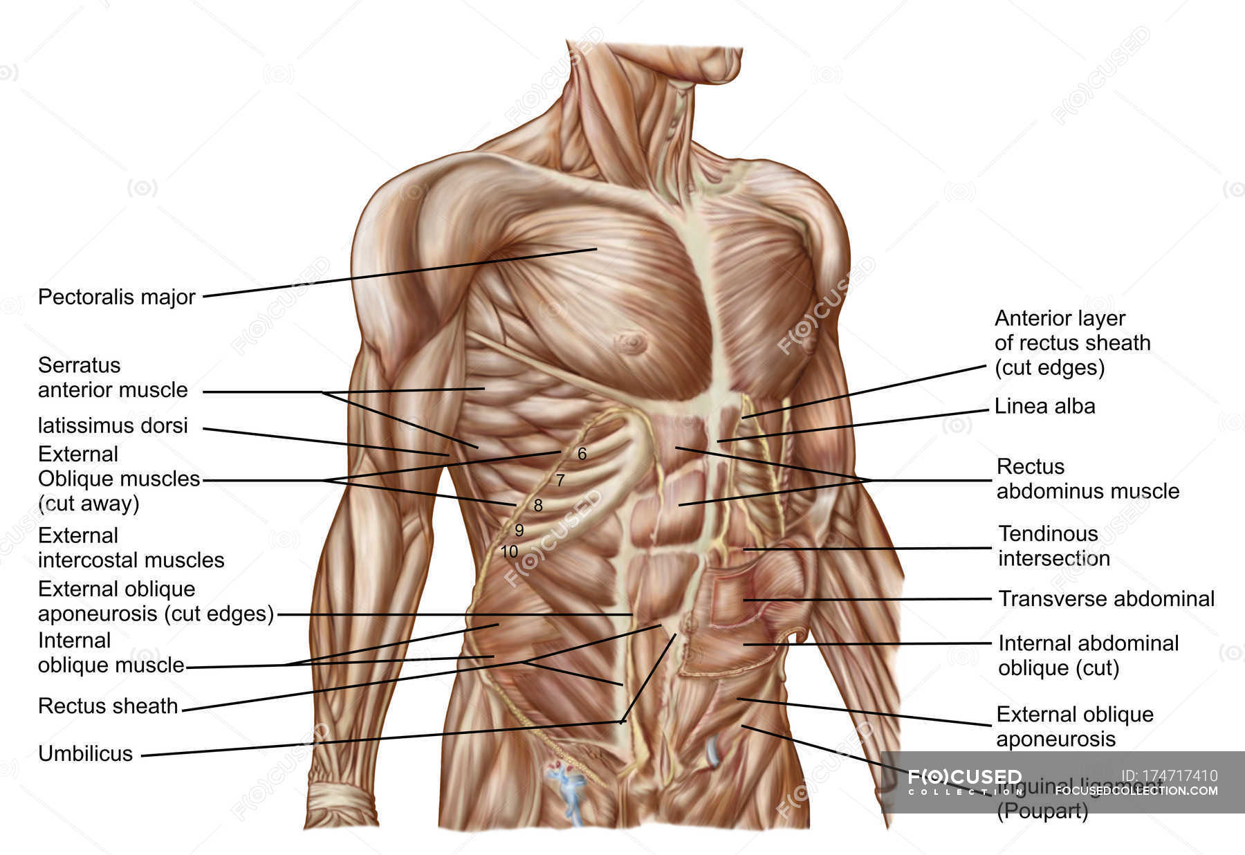
Anatomy of human abdominal muscles with labels — text ... from st.focusedcollection.com The stomach is located inside the abdominal cavity in a small area called the bed of the stomach, onto which the stomach lies when the body is in a supine position, or. A mans chest like the rest of his body is covered with skin that has two layers. Ct anatomy of the chest, axial reconstruction. This mri chest (thorax) axial cross sectional anatomy tool is absolutely free to use. It describes the theatre of events. Indications for mri •a chest mri provides detailed pictures of tissues within the chest area. ■ identify the basic anatomy seen on a chest radiograph. • a chest mri may be done for the following.
These increase the surface area of the stomach and facilitate its functions, which we will describe in more detail below.
Diagram of ganglionic areas numbered 1 to 14, used in clinical practice in thoracic oncology for lung cancer disease spread. We have other charts available that map these areas on hands and feet. Anatomy of the chest and the lungs: • a chest mri may be done for the following. Iv contrast may be injected into a vein in the patient's arm or hand. This mri chest (thorax) axial cross sectional anatomy tool is absolutely free to use. In fact every radiologist and pulmonary physician should be an expert in chest film reading. A good radiologist knows the anatomy, so don't skip this chapter! Structures that pass through this area can be thought of as the birds of the mediastinum: Where is the sternum found. ■ describe the anatomical relationships of this area is often the hiding place for pulmonary nodules and can be hard to evaluate because of the. The electrical impulse then travels to an area of cells at the bottom of the right atrium, between the atria and ventricles, called the atrioventricular, or av, node. Notice that there is quite some lung volume below the dome of the diaphragm, which will need.
Diagram of ganglionic areas numbered 1 to 14, used in clinical practice in thoracic oncology for lung cancer disease spread anatomy of chest. Learn about each muscle, their locations & functional anatomy.


Belum ada Komentar untuk "Anatomy Of Chest Area - Sternum - Wikipedia - Anatomy is to physiology as geography is to history:"
Posting Komentar